date of publication : 2018-11-13 10:28:54
عدد المشاهدات : 224
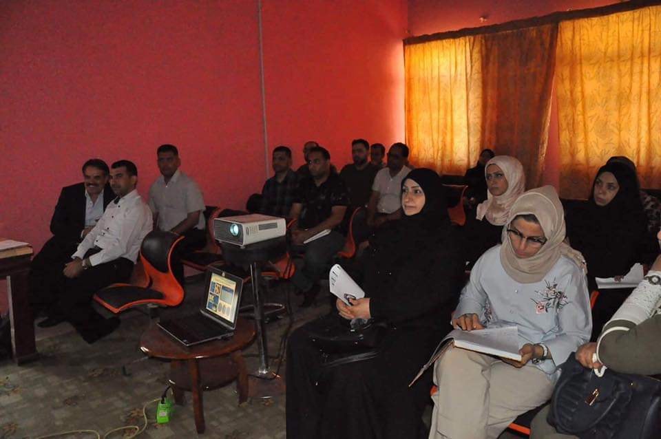
A discussion course was held at the College of Pharmacy, the University of Basrah entitled (Nuclear Medicine Technology).
The researcher, Amer Khazal Al-Hasan illustrate that modern nuclear medicine, which is based on the use of radioactive nuclei with different drug-compounds for the purpose of Physiological photography Testing of the live organs rather than anatomical photography, as well as radionuclides therapy, is an example of thyroid disease and various tumors.
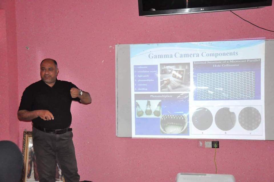
The organization of laboratory preparation and various radionuclides has been explained and the half-life has been done for bone, brain, kidney, liver, lung and heart examinations. Types of tests include static and transportable, CT scans and integrated diagnostics and three-dimensional positron emission. The gamma cameras equipment used in nuclear medicine technology, its main components and the method of work, as well as the tests of quality control, were showed and the pictures that produced in this technique for the tests of bones, thyroid, heart and general organs of the body and methods of diagnosing the disease were reviewed as well.
CT scans require high techniques such as the presence of a sophisticated chemical laboratory and the availability of a spiral nuclear accelerator for the preparation of short radionuclides (half-life) such as irradiated fluorine 18 which can be used with a diabetic derivative for the purpose of clearly imaging the different cancerous areas of the body. Coronary angiography and coronary vascular function can also be performed with high-resolution three-dimensional images to show areas of congestive failure and the Infarction in both of the rest and fatigue situations. More than one radionuclide can be used in the same test to show more than one level of the image by using the latest programs of gamma rays and three-dimension technique.